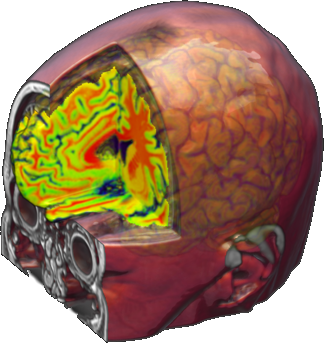
The project H2020 MSCA-RISE 691110 MICROBRADAM is now ending. During its 4-years activities, the Consortium has developed innovative MRI technologies to investigate neurological diseases, and has improved knowledge sharing between leading EU and US institutions.
 Magnetic resonance imaging (MRI) is a main resource for neuroscience and clinical practice. MRI is especially flexible, because the signal can be sensitized to several biophysical and biological phenomena. Importantly, many technological advances can be easily ported to clinical applications.
Microstructural damage is indeed a common feature of many neurological diseases. The MICROBRADAM Consortium developed and validated of a set of advanced microstructural MR techniques for the characterization of tissue damage. The main form of microstructural damage shared by many neurological diseases is related to myelin sheath breakdown. Quantification of myelin is thus critical for the assessment of a variety of neurological diseases including Multiple Sclerosis (MS). The techniques developed have been shown to offer increased specificity and sensitivity to demyelination, compared to conventional approaches.
“In the first half of this project” –says the project coordinator, Federico Giove – “we developed advanced MRI approaches based on non-adiabatic relaxation called RAFFn (Relaxation Along a Fictitious Field of rank n). These are especially sensitive to slow molecular motion that is in turn modulated by myelin damage. We obtained very high correlation between relaxation and myelin content as assessed by quantitative histology in rodents. Applications to humans are straightforward and very promising, especially for Multiple Sclerosis early diagnosis and characterization.”
The Consortium developed functional imaging methods as well, in order to identify the functional correlates of microstructural damage. The project efforts were focalized in two fields: steady state functional connectivity and neuromodulation. Functional connectivity is an approach that quantifies the network behavior of the brain, and is especially attractive for pathologies because it does not assume any specific area of functional response, but characterizes the cortex as a whole. Neuromodulation is important as well, because allows isolating specific neural processes.
“The activities of the Consortium” – concludes Dr. Giove – “have clearly shown that the multiparametric combination of various quantitative techniques has a distinct advantage over the choice of a specific, albeit sensitive approach, especially when a large biological variability is expected.”
The Consortium gathered partners from Finland (University of Eastern Finland and Chares River, Kuopio), Czech Republic (Masaryk University and Clinical Research Center of St. Anne's University Hospital in Brno), Italy (Centro Fermi in Rome and University of Salerno) and US (Center for Magnetic Resonance Research in Minneapolis). The good results and excellent scientific collaboration fostered by the EU H2020 funding will be expanded into technological products and further research projects.