Preliminary results
The Consortium has obtained the first preliminary results, that has been presented at the 25th Annual Meeting of the International Society for Magnetic Resonance in Medicine, in 2017. The results will be published on this website as soon as accepted for presentation.
Results of the first 2 years
In the first half of this project we developed advanced MRI approaches based on non-adiabatic relaxation called RAFFn (Relaxation Along a Fictitious Field of rank n), studying in particular the ranks n=4 and 5. These are especially sensitive to slow molecular motion, expected to be substantially impacted by myelin damage. We measured myelin content and integrity in the normal brain and in complex pathologies in rodents. We obtained very high correlation between relaxation time constants and myelin content as assessed by quantitative histology. Importantly, the correlation we obtained is much improved compared to what can be achieved with conventional MRI approaches. Sensitive detection of demyelination was possible in various brain areas, with different anatomical properties.
In parallel with the development of these advanced structural MRI techniques, we are developing appropriate functional imaging methods, in order to identify the functional correlates of microstructural damage. Our efforts were focalized in two fields: steady state functional connectivity and neuromodulation. Functional connectivity is an approach that quantifies the network behavior of the brain, and is especially attractive for pathologies because it does not assume any specific area of functional response, but characterizes the cortex as a whole. Neuromodulation, that is a byproduct of some of the MRI techniques we developed is important as well, because allow to isolate specific neural processes.
Main results we obtained include a study on semantic network in Alzheimer’s Disease (AD), that showed that in mild AD brain regions belonging to the semantic control network are abnormally connected not only within the network, but also to other areas known to be critical for language processing.
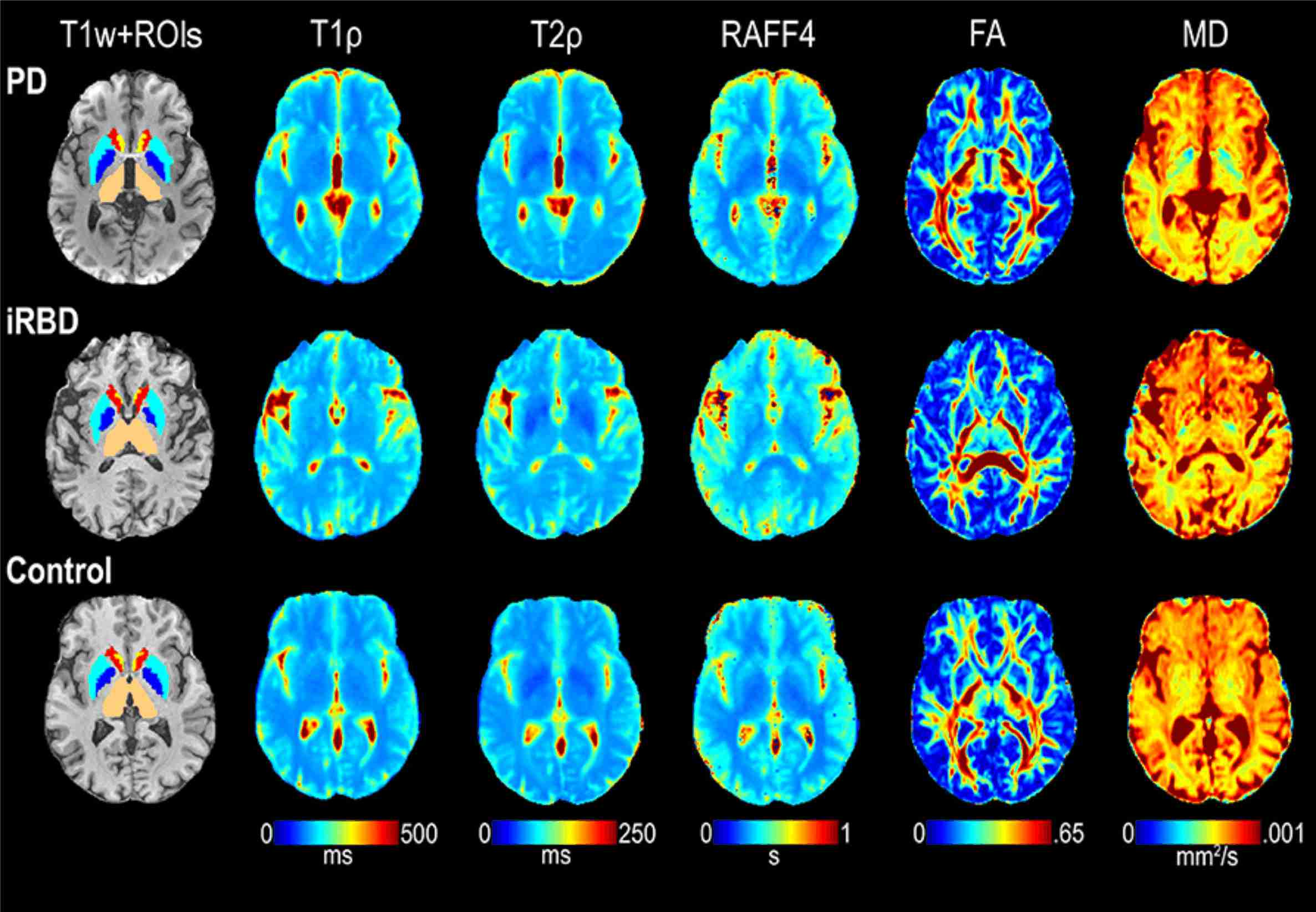
Given the complex features of microstructural damage, it is likely that what is needed is a multiparametric integration of different metrics, where the new approaches we propose are intended to complement other quantitative techniques. MRI in itself lends naturally towards multiparametric studies, because it can produce images sensitized to multiple contrast mechanisms, as described above. MRI can also be combined with compatible approaches, including PET based molecular imaging, electrophysiological measurements and neuromodulation. In this project, we implemented an appropriate set of processing tools to combine different kind of information, for now derived mainly from MRI measurement. Main results obtained with this approach are related to Parkinson Disease (PD) and the associate idiopathic REM sleep behavior disorder (iRBD). We were able to show that rotating frame relaxation methods, along with functional connectivity measures, are valuable to characterize iRBD and PD subjects, and with proper validation in larger cohorts may provide pathological signatures of iRBD and PD.
Impact
The basic effectiveness in PD, MS, iRBD, AD is being directly tested by this study, and unprecedented sensitivity to detect demyelination in vivo and non invasively has been already proven. However, we expect that the systematic transition of the newly developed techniques to human applications in a clinical environment will offer significant advantage also in other pathologies not directly addressed by this project. Therefore, insights gained from this project may ultimately have a significant impact on reducing the morbidity associated with neurodegeneration in general. Any new tools available for early detection and therapy assessment in neurodegeneration has a great potential impact, given the enormous personal and public health burden of neurodegenerative diseases. The tools developed within this project will have the added value of being non-invasive and easily manageable in a clinic environment.
Information obtained from this project is expected to improve patient care through early disease detection and better assessment of disease progression or treatment in the future. In particular, we believe that the information gained in this investigation will be important in developing new ways to monitor for changes in the brain that occur in neurodegeneration in general.
We envisage that this process will happen in two stages. A first stage, that is already occurring, is the diffusion of the advanced methods we are developing to an increasingly wide community of neuroscientists. The availability of such sensitive tools to characterize microstructural brain damage and the relevant functional counterpart is improving the quality of the research of the involved research teams, and we committed ourselves to widen as far as possible the dissemination of our approaches.
In a second, long term stage our techniques (together with hardware improvements of the MRI instrumentation) have the potential to be applied to single patients to improve and personalize the diagnosis and the treatments.